Hot, Swollen Foot Without a Wound (Charcot Foot)
In diabetes, a hot, swollen and often painless foot with no wound may be Charcot foot. Why the first step is to stop walking on it, and how it is managed.
Short answer
In a person with diabetes and nerve damage, a hot, swollen and often painless foot with no wound may be an active Charcot foot. In this condition the bones and joints of the foot become weak, and continuing to walk on it can lead to lasting deformity. The first step is to stop putting weight on the foot and see a doctor within a few days. If there is fever or spreading redness, same-day assessment is needed.
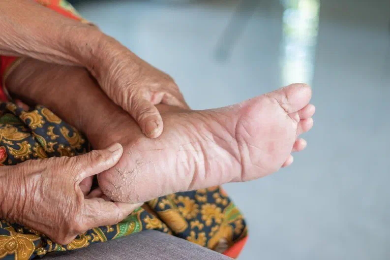
In a person with diabetes and loss of feeling in the feet, one foot that is clearly warmer and more swollen than the other, and sometimes red, although there is no wound, suggests an active Charcot foot. The medical name is Charcot neuro-osteoarthropathy. In this condition an inflammatory process begins in the bones, joints and ligaments of the foot, and the bones become temporarily weak. Because loss of feeling dulls the pain, the person keeps walking, and the weakened bones can shift under load. When it is noticed early and the foot is protected, it is often possible to preserve the shape of the foot.
What can it be mistaken for?
Because Charcot foot is not a common condition, it can be mistaken for other problems at first glance. Guidelines recommend that a hot, swollen foot without a wound in a person with diabetes and nerve damage is treated as a Charcot foot until shown otherwise.
| Condition | Typical features | How it differs from Charcot foot |
|---|---|---|
| Active Charcot foot | Hot, swollen, sometimes red foot, with little or no pain and usually no wound | Fever is usually absent, and redness may fade when the foot is raised |
| Skin and soft tissue infection | Redness, warmth and tenderness, usually starting from a wound or crack | There is an entry point, and fever and raised blood markers may be present |
| Bone infection | A long-standing wound that does not close, with bone involvement beneath | Almost always occurs together with a wound |
| Gout | Sudden, very painful joint swelling, usually at the base of the big toe | Pain is prominent |
| Deep vein thrombosis (clot) | Swelling and tightness in the calf | The swelling is in the leg more than the foot, and Doppler ultrasound tells them apart |
| Sprain or fracture | A history of a fall or strain | In a person with loss of feeling, a minor injury can also set off the Charcot process |
These conditions are treated differently. When a Charcot foot is taken for infection, only antibiotics are given and the person keeps walking, valuable time can be lost. On the other hand, missing a true infection is also harmful. This is why a doctor needs to tell them apart. For a red foot with fever, see the page on swollen, red foot with fever.
Until you see a doctor
- Keep weight off the foot. Use crutches, a walking frame or a wheelchair if you can. This is the most important thing you can do.
- Keep the foot raised when sitting and lying down.
- Do not apply heat or cold, and do not massage the foot.
- Do not start antibiotics, painkillers or water tablets on your own.
- Compare the two feet and take a photo. Compare the warmth of both feet with the back of your hand.
- Look at the sole and between the toes, and note whether there is a wound, crack or blister.
- Measure your temperature and your blood sugar.
What does the assessment include?
- Examination: The two feet are compared. Swelling, redness, changes in shape, the presence of a wound, sensation and pulses are assessed.
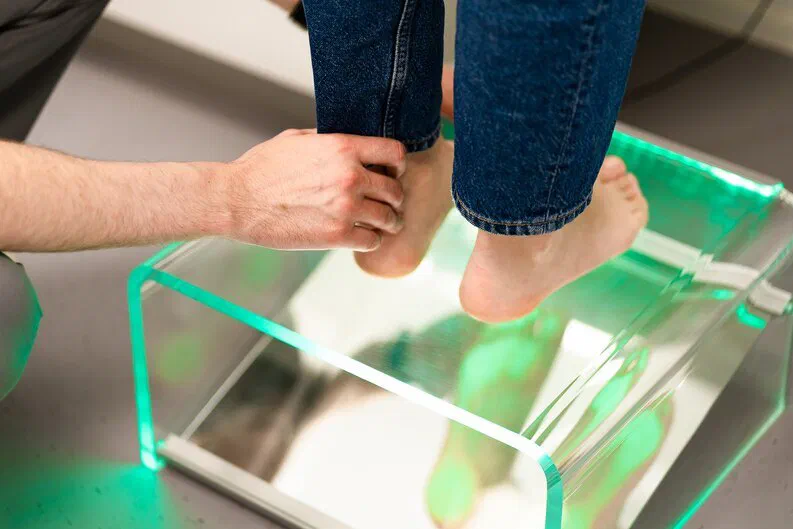
- Temperature measurement: Skin temperature is measured at the same points on both feet with an infrared thermometer. The difference is used both for diagnosis and for follow-up.
- X-ray: This is the first imaging test. It can be normal in the early phase.
- MRI: If the X-ray is normal but suspicion remains, an MRI is requested. It can show early changes in the bone and helps in telling Charcot foot from infection.
- Blood tests: Infection markers, blood sugar and kidney function. In Charcot foot, infection markers are often normal or mildly raised, and they do not make the diagnosis on their own.
Guidelines recommend starting to protect the foot even while the diagnosis is being confirmed. Continuing to walk while waiting for the imaging result is harmful.
How is it treated?
In the active phase, the basis of treatment is not medication. It is keeping weight off the foot and immobilising it.
- Knee-high cast or device: Guidelines recommend immobilising the foot and ankle in a cast that reaches to below the knee and preferably cannot be removed by the patient (a total contact cast), or in a knee-high walking device. This is applied by orthopaedics and teams experienced in this field.
- Regular checks: The cast is changed at regular intervals, the skin is checked and the temperature difference is measured. The cast is renewed as the swelling goes down.
- When is it stopped? The active phase is considered to have settled when the temperature difference between the two feet has gone, the swelling has reduced and imaging shows that the bones have settled. This usually takes months.
- Afterwards: The return to walking is gradual. Custom shoes and insoles that fit the shape of the foot help prevent a flare-up and the development of wounds.
- Surgery: If there is marked deformity, an unstable joint or a recurring wound caused by a prominent bone, orthopaedics may consider surgical options.
- The other foot: The other foot, which carries the load, should also be checked regularly.
Blood sugar control and general foot care continue throughout. No medicine has been shown to stop the Charcot process, and guidelines do not routinely recommend medication for this purpose.
Assessment in Istanbul
The first question to answer in a hot, swollen foot is whether this is infection or Charcot foot, and the two can also occur together. Prof. Dr. Aytaç Çetinkaya, a specialist in Infectious Diseases and Clinical Microbiology, assesses the infection side of this distinction, helps avoid unnecessary antibiotic use and, if Charcot foot is suspected, recommends working with orthopaedics for casting and follow-up. If there are no urgent signs, you can send a comparative photo of both feet, along with any X-ray and MRI reports, via WhatsApp for a preliminary review. A preliminary review does not replace an examination. Keep weight off the foot while you wait for a reply.
Frequently asked questions
What is Charcot foot?
I have no pain. Can it still be serious?
My X-ray was normal. Does that rule out Charcot foot?
How long is the cast or boot needed?
Are antibiotics needed for Charcot foot?
References
- IWGDF Guidelines on the diagnosis and treatment of active Charcot neuro-osteoarthropathy in persons with diabetes mellitus (2023)
- IWGDF/IDSA Guidelines on the diagnosis and treatment of diabetes-related foot infections (2023)
- IWGDF Practical guidelines on the prevention and management of diabetes-related foot disease (2023)
This page is for general information only and does not replace an examination, diagnosis or treatment. In an emergency, call your local emergency number or go to the nearest emergency department.